€700,000 boost for preeclampsia research

Two percent of all pregnant women in Europe develop preeclampsia – a condition that occurs after the 20th week of pregnancy and causes sudden high blood pressure and organ dysfunction. The elevated pressure in the blood vessels can damage both the mother’s blood vessels and those in the placenta. As a result, the unborn baby no longer receives sufficient nutrients and oxygen. This may lead to growth problems, long-term damage, or even miscarriage or stillbirth. In addition, the mother has a much higher risk of developing cardiovascular disease later in life.
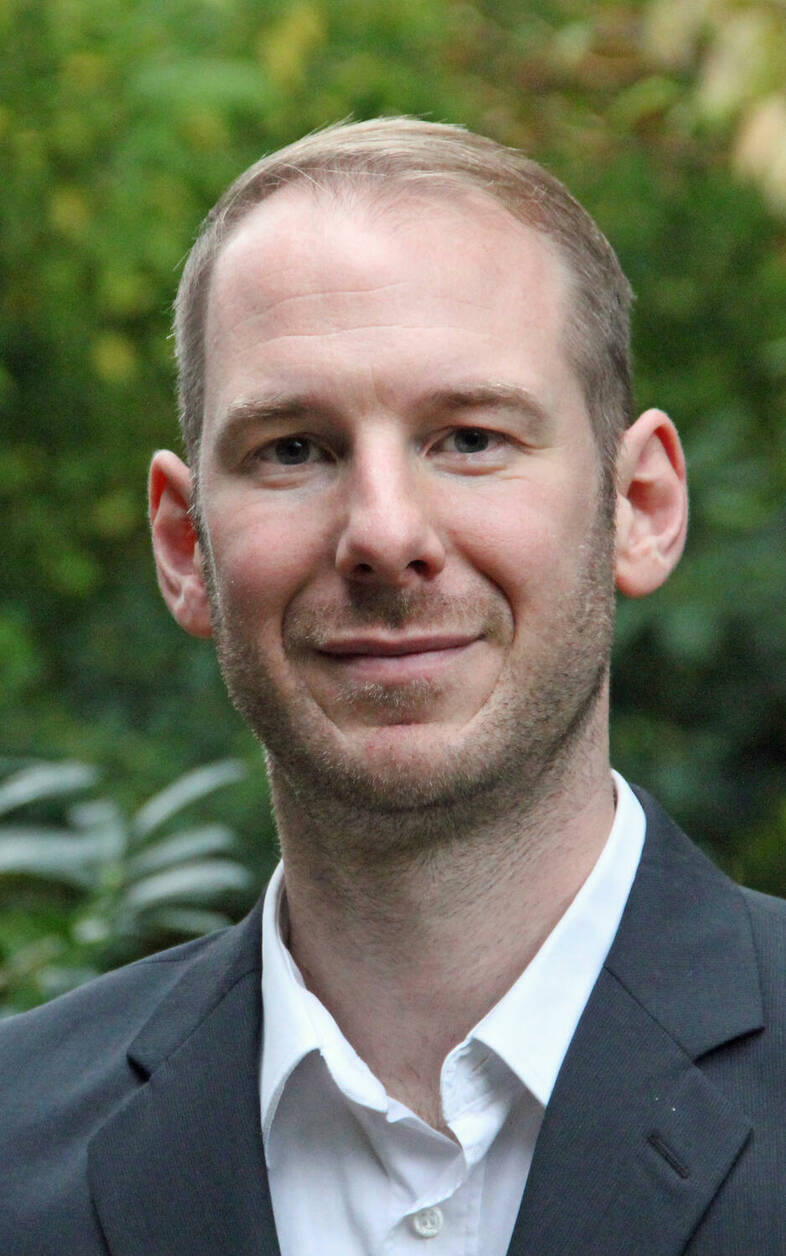
The causes and mechanisms of the disease are still not fully understood, but there is no doubt that the placenta – and its Hofbauer cells in particular – play a crucial role. Hofbauer cells are macrophages, a type of white blood cell that roams the body and kills pathogens. “Studies using animal models have shown that the clinical symptoms of preeclampsia disappear immediately after removal of the placenta,” explains Dr. Florian Herse. He is a researcher in Professors Dominik N. Müller’s and Ralf Dechend’s Hypertension-Mediated End-Organ Damage Lab at the Experimental and Clinical Research Center (ECRC), a joint institution of the Max Delbrück Center and Charité – Universitätsmedizin Berlin.
Together with Professor Martin Gauster of the Medical University of Graz, Herse plans to further investigate the development of these macrophages and their dysregulation in preeclampsia. The two researchers have been collaborating for ten years and will soon receive a D-A-CH grant for the second time. The funding process enables research proposals to be jointly submitted by the German Research Foundation (DFG) and its partner organizations, the Austrian Science Fund (FWF) and the Swiss National Science Foundation (SNSF). Of the €700,000 in total funding, €372,000 will go to the Max Delbrück Center.
Shedding light on placenta impairment
Gauster and his team have developed a flow culture model for placental tissue that is capable of simulating the various states of health during pregnancy. In Berlin, Herse and Dr. Fabian Coscia’s lab are using single-cell sequencing and microscopy-guided proteomics to examine and compare the spatial and temporal dynamics of macrophage development in healthy and diseased placental tissue.
Hofbauer cells don’t circulate freely through the blood vessels but remain in the fetal area of the placenta, where they appear to prevent pathogens from passing from the mother’s blood to the unborn child. The number of these cells is reduced in pregnancies with severe preeclampsia. “We suspect that this changes the structure of the blood vessels and that of the tissues surrounding them, thereby impairing the function of the placenta,” says Herse. “So now we want to examine more closely how the macrophages and blood vessels interact.”
Text: Catarina Pietschmann
Further information
- Müller/Dechend Lab, Hypertension-caused End-Organ Damage
- Coscia Lab, Spatial Proteomics
- Research Team Gauster, Research Focus: Reproduction, Pregnancy and Regeneration


